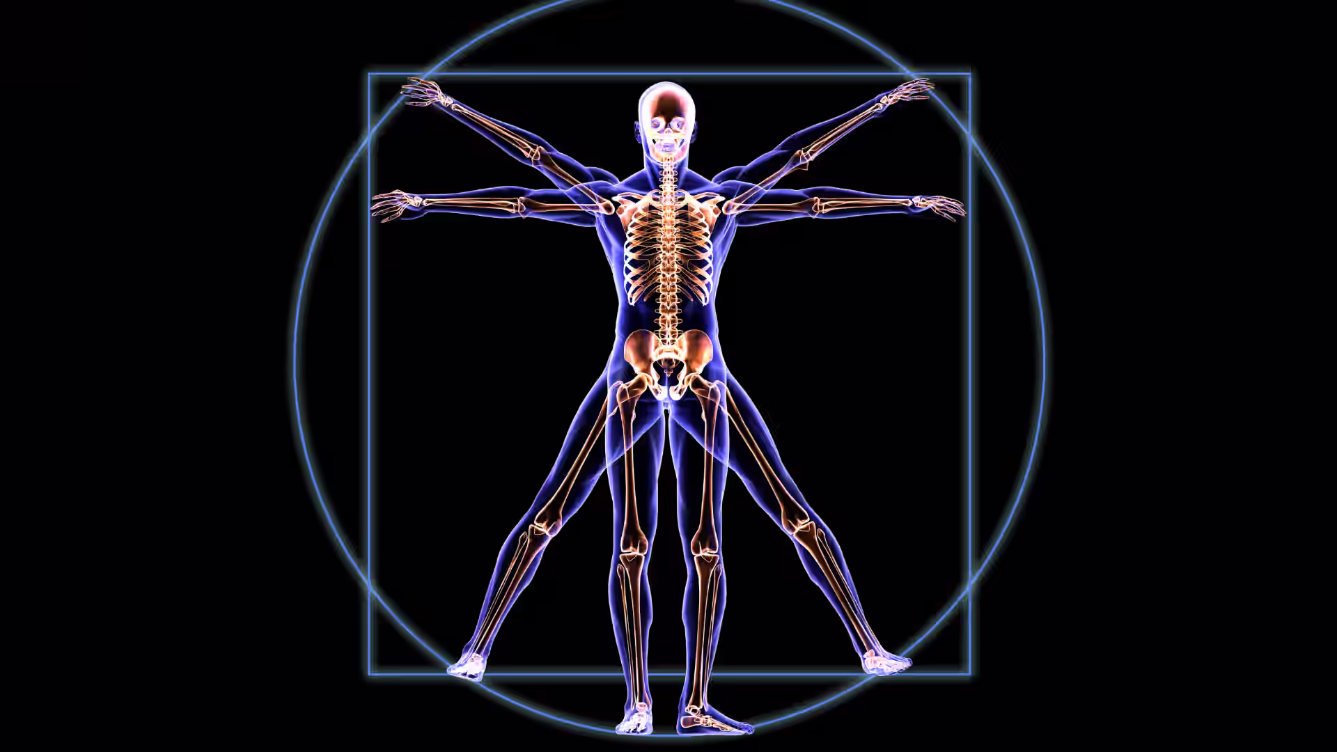


Often referred to as a "sunken chest" or “funnel chest,” pectus excavatum is more than a cosmetic issue—it can affect the heart and lung function, especially in moderate to severe cases.
The most common congenital chest wall deformity, pectus excavatum, affects approximately 1 in every 400 individuals, more frequently in males than females. Thanks to medical advances, many people with pectus excavatum now have access to effective, evidence-based treatments that can improve appearance and health outcomes.
This guide explains pectus excavatum, its causes, the symptoms to look out for, and the full range of treatment and recovery options available today.
[signup]
Understanding Pectus Excavatum
Before reviewing the effects and treatments of pectus excavatum, it helps to understand what the condition actually involves.
What Is Pectus Excavatum?
Pectus excavatum occurs when the cartilage that connects the ribs to the sternum grows abnormally, causing the sternum to sink into the chest cavity. This creates a concave appearance that can range from mild to very deep. The condition can become more pronounced during periods of rapid growth, such as puberty.
How Common Is It?
- Occurs in roughly 1 in 300 to 1000 live births
- It is more common in males (about 5 times more likely than in females)
- Often becomes noticeable in early childhood and typically worsens in adolescence
Symptoms and Complications
The signs and symptoms of pectus excavatum vary greatly depending on severity.
Recognizing Symptoms
Mild cases may have no symptoms beyond the visual indentation. In moderate to severe cases, individuals may report:
- Chest tightness or discomfort
- Reduced endurance, especially during aerobic activities like running
- Rapid heartbeat or palpitations
- Difficulty taking deep breaths These symptoms are often more noticeable during exercise or physical exertion.
Complications
More severe defects can impact the heart, lungs, and pulmonary function.
Heart and Lung Impact
A deeply sunken chest may push the heart to the left, compressing it and limiting its ability to pump blood efficiently. The lungs may also be restricted, reducing lung volume and airflow.
Psychological Impact
Self-consciousness is common, especially in teens. This may lead to:
- Avoiding sports or swimming
- Wearing baggy clothes
- Social withdrawal or anxiety
Causes and Risk Factors
Understanding the root causes of pectus excavatum helps guide diagnosis and treatment.
Genetic Links
Pectus excavatum sometimes runs in families and is more common in people with connective tissue disorders:
Environmental Factors
While the exact role of environmental influences remains unclear, researchers continue to explore whether in-utero development factors, nutritional status, or mechanical pressure in infancy could play a role.
Risk Factors by Age Group
- Infants may show signs at birth, though many cases are mild early on.
- Children and teens often experience worsening symptoms during growth spurts.
- Over time, adults with untreated cases may experience increased fatigue, back pain, and respiratory symptoms.
Diagnosis
Diagnosis begins with a physical exam and is confirmed with imaging and function tests.
Physical Examination
A healthcare provider may assess the following:
- The depth and symmetry of the chest depression
- Spinal alignment (as scoliosis may co-occur)
- Breathing pattern and chest expansion
Imaging Tests
- Chest X-ray: Reveals the shape of the sternum and ribs
- CT scan: Provides precise measurements and helps calculate the Haller index
- MRI (occasionally used): Offers more detail for soft tissue and heart involvement
Pulmonary Function Tests (PFTs)
Pulmonary function tests (PFTs) assess how well the lungs work and determine whether the sunken chest interferes with breathing. These tests are especially useful when patients report symptoms like shortness of breath, fatigue during exercise, or chest tightness.
Commonly referenced PFT values include:
- Forced Vital Capacity (FVC): The total amount of air exhaled during a forceful breath out. A reduced FVC may suggest restricted lung expansion due to chest wall compression.
- Forced Expiratory Volume in 1 Second (FEV1): The amount of air exhaled in the first second of a forceful breath. This can help detect airway obstruction or restriction.
- FEV1/FVC Ratio: Helps differentiate between obstructive and restrictive lung conditions.
- Total Lung Capacity (TLC): This may be measured in some cases to evaluate how much air the lungs can hold at maximum inflation.
In patients with pectus excavatum, PFT results may show a mild restrictive pattern, especially if the chest indentation is severe and affects lung expansion.
Haller Index
The Haller index is a measurement used to assess the severity of pectus excavatum based on chest CT scans.
- Calculated by dividing the chest’s width by the depth of the chest cavity
- A value >2.5 is typically considered a threshold for surgical evaluation
Differential Diagnosis
- Ehlers-Danlos syndrome
- Marfan syndrome
- Noonan syndrome
- Scoliosis
Treatment Options
Treatment depends on age, severity, symptoms, and personal preferences.
Non-Surgical Treatments
- A non-invasive option that uses suction to slowly lift the chest wall
- Best for younger patients with flexible bones
- Requires commitment to daily use (up to several hours/day for months or years)
Posture Correction
- Exercises to improve spinal alignment and open the chest
- Includes back extensions, chest-opening stretches, and shoulder blade strengthening
Breathing Training
- Resistance devices or techniques to strengthen respiratory muscles and improve lung capacity
Surgical Treatments
- Small incisions are used to insert a metal bar beneath the sternum
- The bar remains in place for 2–4 years to reshape the chest wall
- Most common approach for children and teens
- Involves the removal of abnormal cartilage and sternal repositioning
- Typically recommended for older patients or severe, asymmetrical cases
Emerging Techniques
Magnetic Mini-Mover Procedure (MMPP): This procedure uses magnets to pull the sternum forward over time. It is still under investigation and not widely available.
Recovery and Prognosis
Recovering from pectus excavatum treatment—whether surgical or non-surgical—requires careful planning, follow-up, and support.
Post-Treatment Care
After a procedure such as the Nuss or Ravitch surgery, patients typically stay in the hospital for a few days for monitoring and pain management. Recovery continues at home with the following:
- Restricted physical activity for 4–6 weeks to allow proper healing of the chest wall
- Avoidance of contact sports or heavy lifting for several months
- Post-operative checkups to assess healing and ensure the chest bar (if present) remains in place
For non-surgical treatments like the vacuum bell, recovery is gradual and requires consistent daily use of the device, often for months or even years.
Pain Management Innovations
Pain control is a key part of recovery, particularly after surgical repair. Traditional pain management may involve oral medications, but newer techniques include:
- Cryoablation (nerve freezing): Often done during surgery to reduce post-op pain for several weeks
- Epidural or nerve blocks: Used in some centers to minimize the need for opioids and promote faster mobility
- Non-opioid medications like acetaminophen and NSAIDs as part of a multimodal pain regimen
Physical Therapy Guidelines
Rehabilitation after treatment typically includes:
- Breathing exercises to improve lung capacity
- Posture training to prevent the recurrence of chest wall collapse
- Light aerobic activity (like walking) introduced gradually under medical guidance
- For surgical patients, physical therapy may begin 4–6 weeks post-op and continue for several months, depending on recovery progress.
Consistent movement supports better healing, lung expansion, and muscle strength when approved by a healthcare provider.
Long-Term Outcomes
Most patients experience:
- Improved chest appearance and posture
- Better exercise tolerance and endurance
- Relief from symptoms such as fatigue and shortness of breath
- Improvement in cardiovascular function
- Enhanced confidence and emotional well-being
For surgical patients, chest bars placed during the Nuss procedure are usually removed after 2–4 years, depending on the patient’s age, bone flexibility, and healing progress.
While recurrence is rare, regular follow-up appointments help monitor for any structural changes or complications. The outlook is excellent with appropriate care, and most individuals return to full activity and a normal lifestyle.
Living with Pectus Excavatum
While treatment can address the physical aspects of pectus excavatum, it’s equally important to support the emotional and social well-being of individuals living with the condition—both before and after treatment.
Coping Mechanisms for Patients and Families
- Support groups can offer connection with others navigating similar challenges.
- Counseling may help with body image concerns or anxiety
- Encourage open communication with schools, coaches, and peers
When to Seek Medical Advice
- New or worsening symptoms (e.g., chest pain, fatigue, shortness of breath)
- Emotional distress or withdrawal due to body image
- Concerns about treatment options or monitoring
[signup]
Key Takeaways
- Pectus excavatum is a manageable condition with proper diagnosis and treatment options tailored to individual needs.
- Pectus excavatum is the most common chest wall deformity, affecting roughly 1 in 400 individuals, and can range from a mild cosmetic issue to a condition that impacts heart and lung function.
- Symptoms vary by severity and may include shortness of breath, reduced exercise tolerance, chest pain, or psychological distress due to body image concerns.
- Diagnosis involves physical exams, imaging (like CT scans), pulmonary function tests, and calculation of the Haller Index to assess severity and guide treatment decisions.
- Non-surgical treatments such as vacuum bell therapy, posture exercises, and breathing resistance training are often used in milder cases or growing children.
- Surgical options, like the minimally invasive Nuss procedure or the more extensive Ravitch technique, offer long-term correction for more severe deformities.
- Recovery includes activity restrictions, pain management innovations (e.g., cryoablation), and physical therapy. Most patients report improved appearance, function, and self-confidence.