


Imagine slicing through the human body like a skilled sushi chef—this is essentially what anatomical planes allow us to visualize.
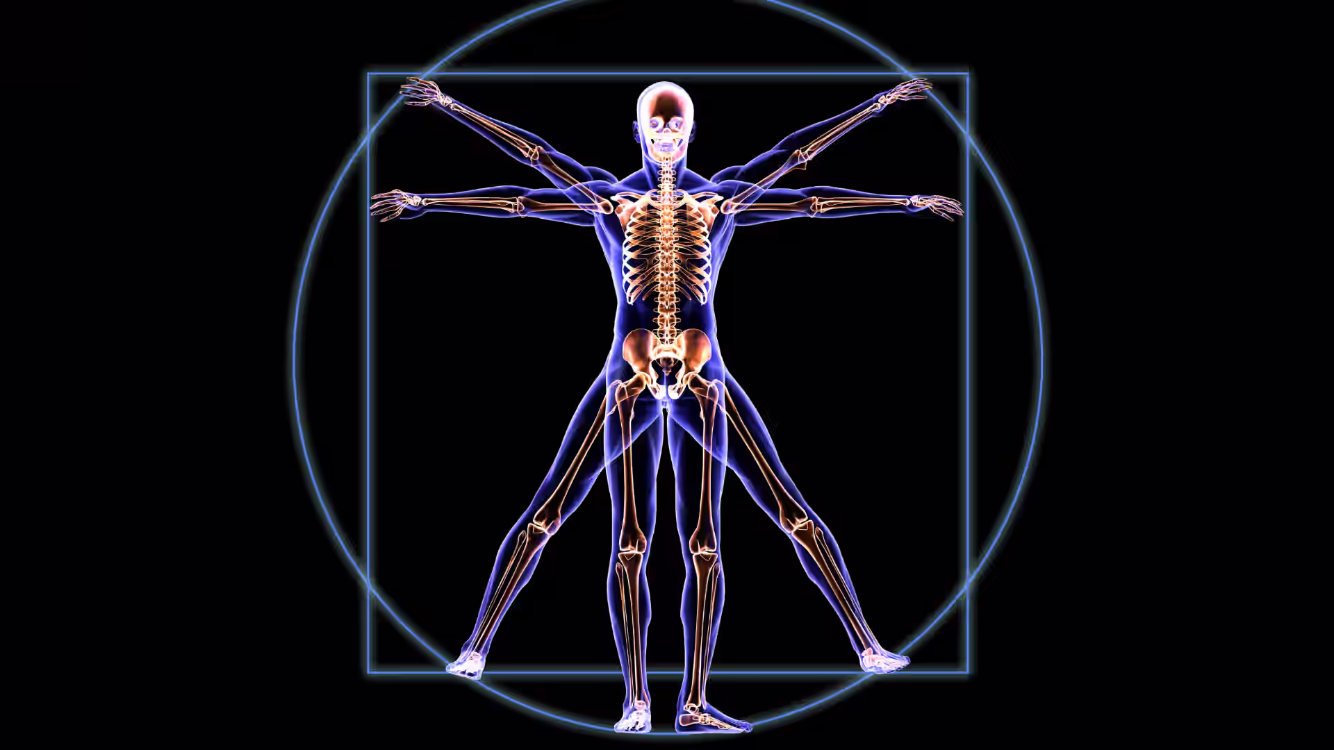
Anatomical planes and directional terms form the foundation of understanding and describing human anatomy. These standardized terms and concepts help medical professionals, students, and anyone interested in the human body explain the body's structure, orientation, and relative positions of organs and tissues.
[signup]
The Fundamentals of Body Planes
Anatomical body planes are imaginary flat surfaces that divide the human body into sections to describe and locate structures within the body when it is stationary and in motion.
The Three Primary Anatomical Planes
The sagittal (lateral) plane divides the body vertically, separating into left and right sides. The midsagittal (median) plane is a specific sagittal plane that passes through the body's midline, cutting it into two equal halves.
The frontal (coronal) plane also separates the body vertically, but divides it into front and back sections.
The transverse (axial) plane divides the body horizontally, separating it into upper and lower halves.
Anatomical Directions and Positional Terms
Before going further, it's important to define anatomical position, as it is the consistent starting point for understanding and describing the body's orientation.
Standard anatomical position is the reference point used in anatomy to describe body parts and their positions. In this position, the body stands upright, facing forward, with the arms at the sides and the palms facing forward. The feet are flat on the ground and parallel to one another, and the body is not rotated.
Directional Terms in Anatomy
Anatomical directional terms are standardized terms used to describe the location or position of structures in relation to other parts of the body.
- Anterior and Posterior: Refer to the front and back of the body, respectively. For example, the kneecap is on the anterior aspect of the leg, while the buttocks are on the posterior aspect of the body.
- Superior and Inferior: Describe the vertical position of body structures. Superior means toward the head and inferior means toward the feet. For instance, the head is superior to the neck.
- Medial and Lateral: Medial refers to structures closer to the midline of the body, while lateral describes structures further away from the midline. For example, the big toe is on the medial aspect of the foot, while the pinky toe is on the lateral aspect.
- Proximal and Distal: Describe the relative position of limbs and appendages. Proximal means closer to the point of attachment to the body, while distal means farther away. For example, the shoulder is located at the proximal aspect of the arm, while the hand is situated distally.
Other Important Positional Terms
- Superficial and Deep: Describe the relative depth of structures within the body. Superficial refers to structures closer to the skin's surface, while deep refers to those further from the surface. The skin is superficial to the muscles, and the muscles are deep to the skin.
- Parietal and Visceral: Refer to the layers of membranes that cover body cavities and organs. Parietal refers to the outer layer, and visceral refers to the inner layer that covers organs. For instance, the parietal pleura lines the chest cavity, while the visceral pleura covers the lungs.
Body Cavities and Regions
The body cavities are spaces that contain internal organs. The body is divided into two main cavities: the ventral and dorsal cavities.
Ventral Cavity
Located at the front of the body, the ventral cavity is further divided into two portions.
The thoracic (chest) cavity houses the heart, lungs, trachea, esophagus, large blood vessels, and nerves. It is bound by the ribs and the diaphragm.
The abdominopelvic cavity is the larger portion of the ventral cavity and can be further divided into two parts:
- The abdominal cavity contains most of the gastrointestinal tract, the kidneys, and the adrenal glands. It is bound by the diaphragm, the body wall, and the pelvic cavity.
- The pelvic cavity contains the bladder, urethra, reproductive organs, and rectum. It is bound by the abdominal cavity, the sacrum, and the pelvis.
Dorsal Cavity
The dorsal cavity is the smaller of the two main body cavities and is located at the back of the body. It is subdivided into the cranial cavity (containing the brain) and the spinal cavity (containing the spinal cord).
Abdominal Regions and Quadrants
The abdomen can be divided into four quadrants or nine regions to facilitate the identification, description, and localization of organs and symptoms.
Abdominal Quadrants
The abdomen can be divided into four quadrants by two imaginary lines: one vertical and one horizontal intersecting at the belly button. These quadrants are:
- Right Upper Quadrant (RUQ): Contains the liver, gallbladder, right kidney, the head of the pancreas, and portions of the small intestine and colon
- Left Upper Quadrant (LUQ): Contains the stomach, spleen, pancreas, left kidney, and portions of the small intestine and colon
- Right Lower Quadrant (RLQ): Contains the appendix, right ovary and fallopian tube (in females), right ureter, and portions of the small intestine and colon
- Left Lower Quadrant (LLQ): Contains the left ovary and fallopian tube (in females), left ureter, and portions of the small intestine and colon
Abdominal Regions
The abdomen can also be divided into nine regions, which are more specific and more precisely localize organs and conditions. These regions are formed by two sagittal and two transverse planes.
- Right Hypochondriac: Contains the liver, gallbladder, right kidney, and portions of the small and large intestines
- Epigastric: Contains the stomach, liver, pancreas, adrenal glands, and portions of the small and large intestines
- Left Hypochondriac: Contains the spleen, part of the stomach, pancreas, left kidney, and portions of the small and large intestines
- Right Lumbar: Contains the ascending colon, right kidney, liver, gallbladder, and small intestine
- Umbilical: Contains the navel, small intestine, transverse colon, and the bottom portions of both kidneys
- Left Lumbar: Contains the descending colon, the left kidney, and part of the spleen
- Right Inguinal: Contains the appendix and cecum
- Pubic: Contains the bladder, sigmoid colon, rectum, and reproductive organs
- Left Inguinal: Contains part of the descending colon and the sigmoid colon
Practical Applications of Anatomical Planes and Directions
In exercise science, body planes help us analyze movement patterns. Specific movements occur along specific planes, allowing physiotherapists and trainers to design exercises that enhance strength, flexibility, and mobility. For example:
- Moving limbs away (abduction) or toward (adduction) the body's midline occurs in the frontal plane.
- Bending (flexion) and straightening (extension) joints occur in the sagittal plane.
- Turning your head right and left (rotation) occurs in the transverse plane.
Body planes also provide standardized reference points for medical imaging, such as MRI, CT, and PET scans. These scans produce two-dimensional cross-sections of the three-dimensional body, so understanding anatomical planes allows for accurate interpretation, diagnosis, and treatment planning.
Clinically, anatomical terminology allows healthcare professionals to communicate effectively and precisely about the locations of symptoms, injuries, and health conditions. Surgeons use anatomical planes and abdominal regions to plan and execute procedures.
Frequently Asked Questions (FAQs)
Why are anatomical planes important in medicine?
Anatomical planes standardize communication among medical professionals, ensuring everyone uses the same references when describing body structures and their relationships.
How do body planes relate to medical imaging techniques?
Medical imaging techniques, such as CT scans and MRIs, produce images based on these anatomical planes, allowing for clear cross-sectional views of the body.
What's the difference between the sagittal and parasagittal planes?
The parasagittal plane is a type of sagittal plane that divides the body into unequal left and right portions.
How are anatomical directions used in describing patient positioning?
Directional terms like anterior, posterior, superior, and inferior describe patients' positioning and physical exam findings, helping healthcare professionals clearly explain the body's orientation during examinations or treatments.
Can you explain the relationship between body planes and movement analysis?
Movements in the body are analyzed along specific planes. For example, flexion and extension occur in the sagittal plane, while abduction and adduction happen in the frontal plane.
[signup]
Key Takeaways
- Anatomical terminology includes directional terms, body planes, and body cavities.
- The understanding of anatomical planes and directional terms is fundamental to the practice of medicine and human anatomy.
- By defining body positions and spatial relationships in a standardized way, medical professionals can communicate clearly, avoid confusion, and accurately describe the human body's structure and function.
- These concepts form the basis for a more detailed study of the body's complex systems and are essential for medical imaging, surgical procedures, and clinical practice.